肌周细胞瘤
Myopericytoma
概述:
是一种位于皮下由卵圆形至梭形的肌样细胞所组成的良性肿瘤,瘤细胞常呈同心圆状围绕血管生长,与肌纤维瘤、血管平滑肌瘤以及血管球瘤构成瘤谱。
发病部位: 四肢、颈部和胸椎
诊断要点:
最常见于中年人,好发于四肢、颈部和胸椎,表现为皮下缓慢性生长的无痛性结节;
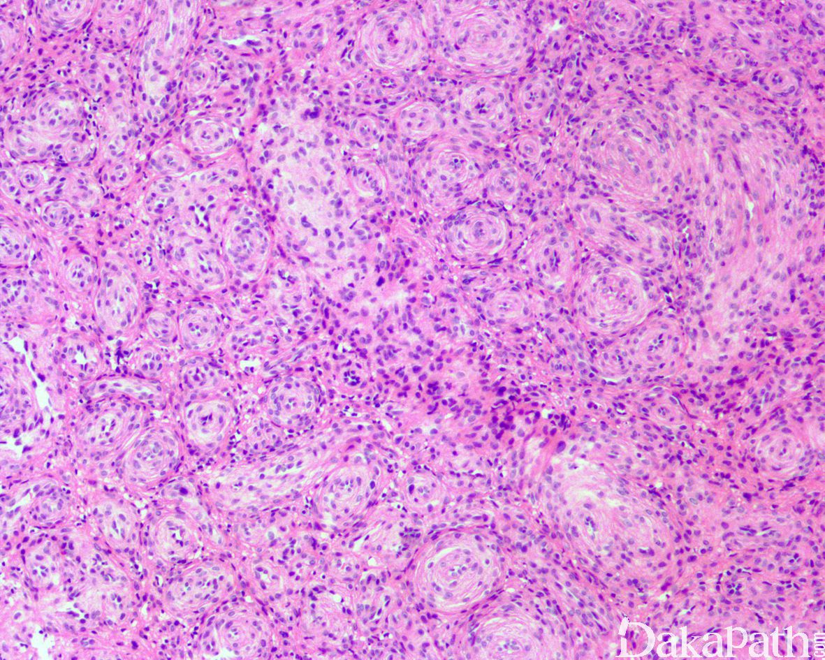
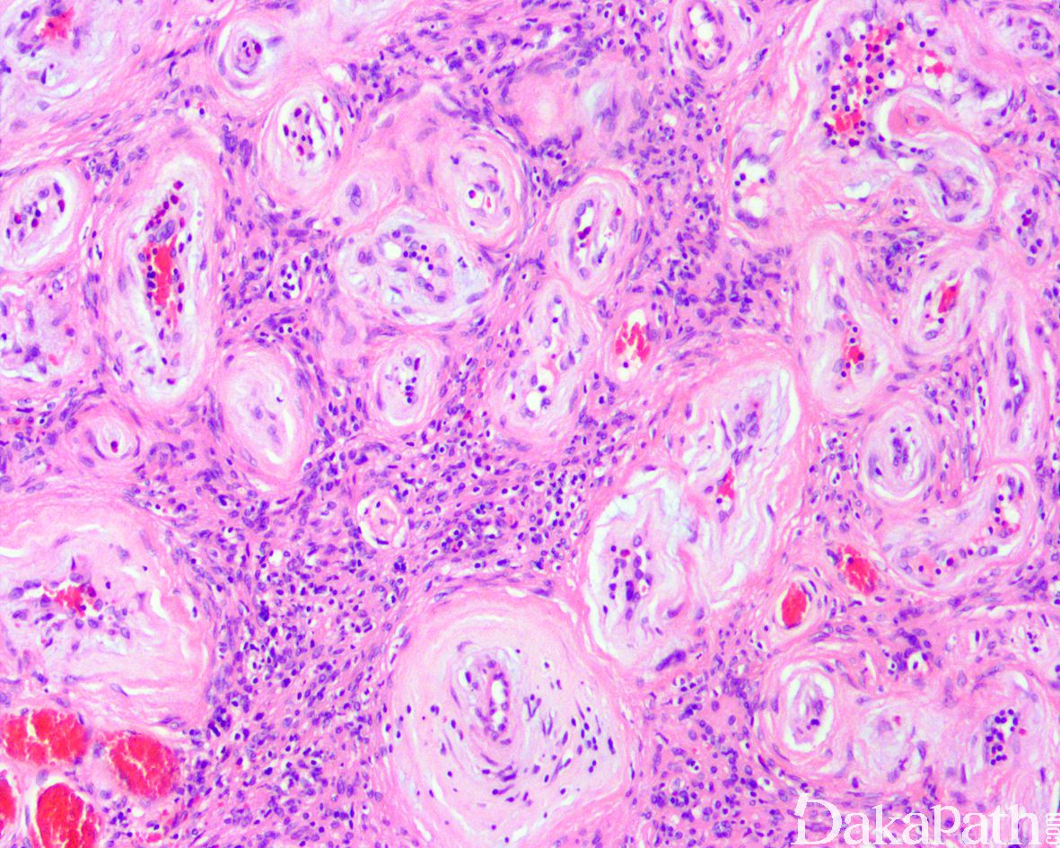
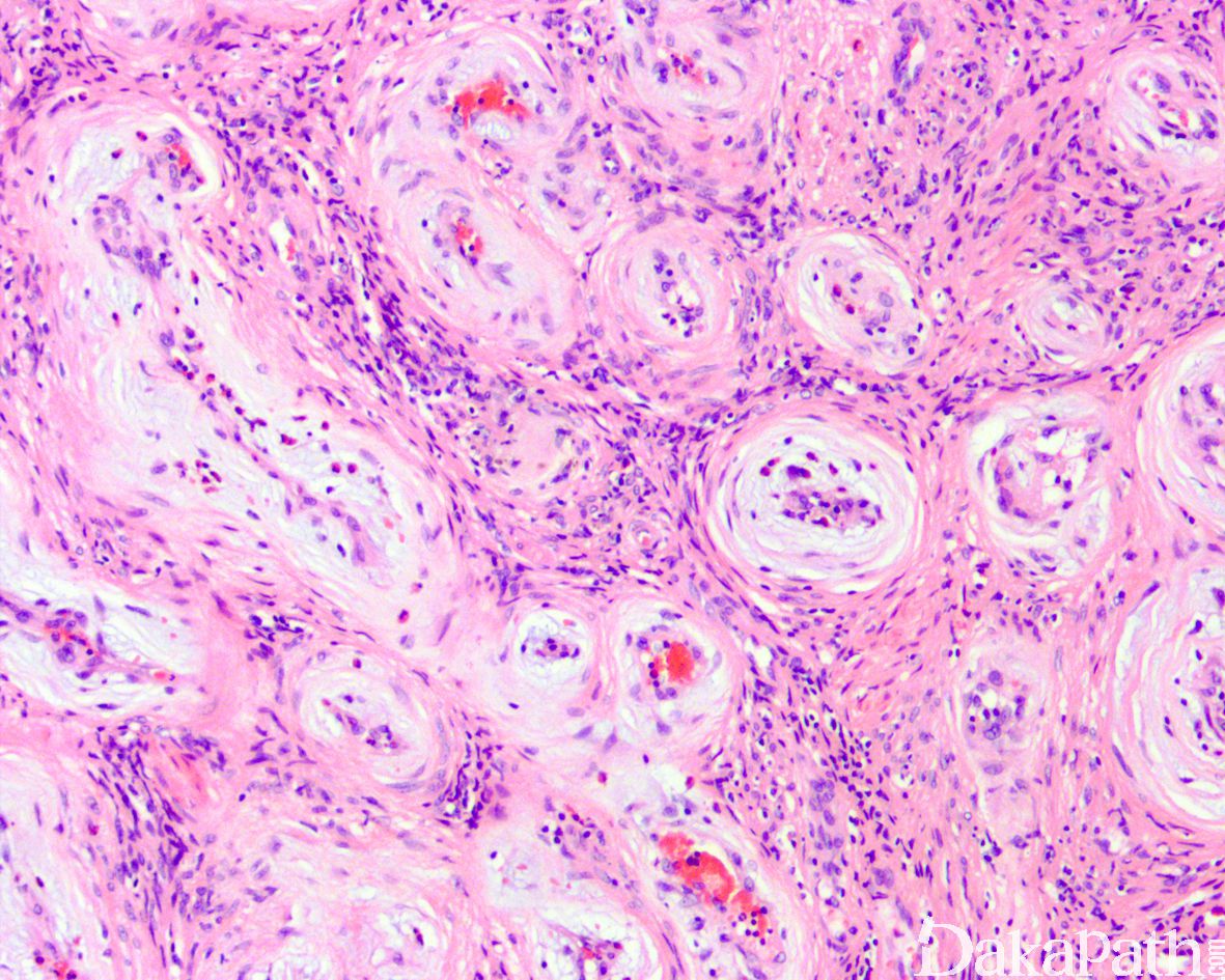
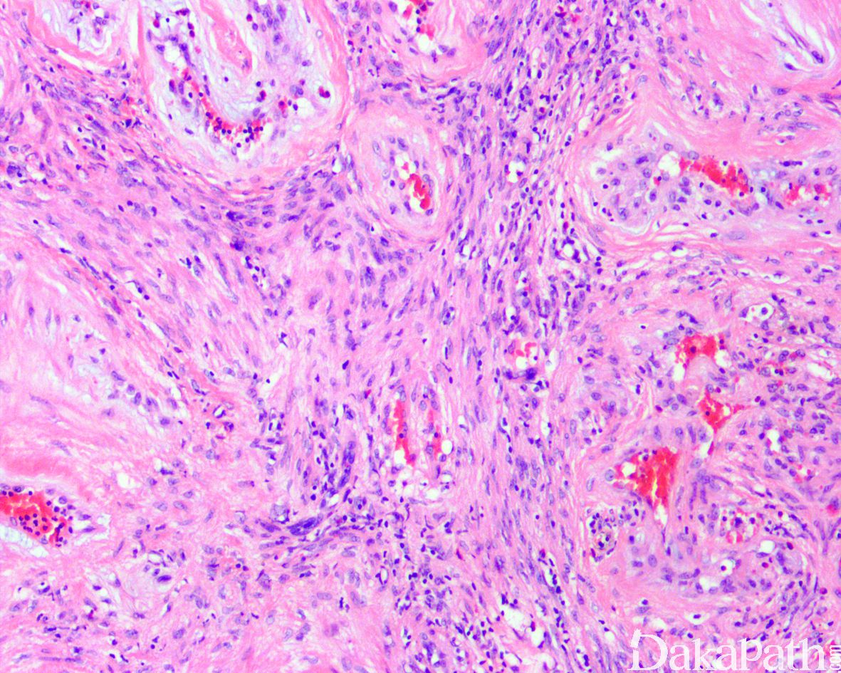
镜下可见形态相对一致的卵圆形至梭形肌样细胞组成围绕小至中等大血管周围呈同心圆状或漩涡状生长;
肌样细胞胞质嗜酸性,核染色质均匀,核异型性不明显,核分裂像一般少于 1/10HPF;
同心圆或漩涡状结构之间基质可伴有黏液样变性;
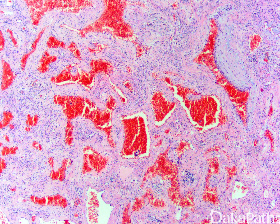
部分病例瘤细胞偏丰富,呈片状分布;局部区域血管也可呈分支状,类似血管外皮瘤;
部分病例内尚可见嗜伊红色、漩涡状的肌样结节,可突向血管腔,类似肌纤维瘤/肌纤维瘤病;
部分病例瘤细胞相对稀疏,间质常伴有玻璃样变性;
少数病例肿瘤完全位于静脉性血管腔内;
少数病例可发生于 AIDS 患者并伴有 EBV 感染, 常表现为多发,多见于较为特殊的部位如颅内,椎管内,支气管等部位,组织学上瘤细胞较丰富,可呈上皮样特征,可见核分裂象,背景中可见较多的淋巴细胞浸润;

免疫组织化学染色:
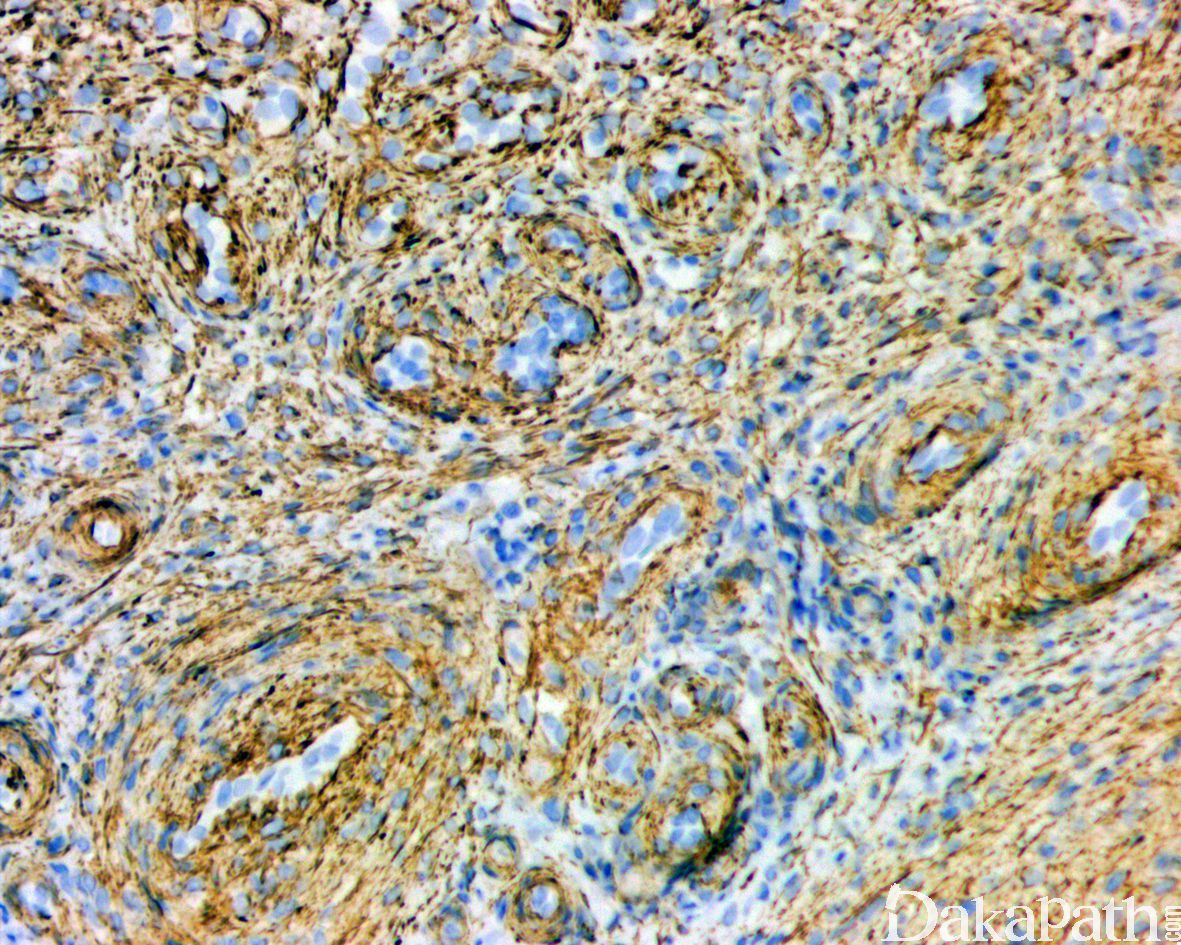
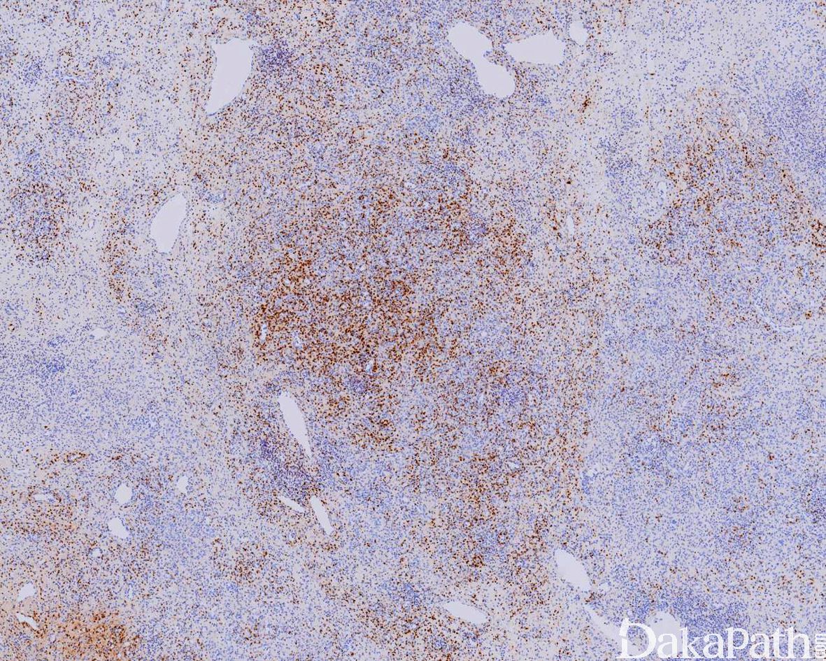
瘤细胞 α-SMA 常弥漫阳性,desmin 和 CD34 偶尔阳性,S-100 蛋白和 CK 阴性。
分子标记:
部分病例显示 t(7;12)(p22;q13),导致 ATCB-GLI1 融合
鉴别诊断:
肌纤维瘤:好发于 2 岁以内的婴幼儿,常多结节生长,有独特的双相表现,常伴黏液变性的间质。
血管平滑肌瘤:皮下深层的痛性小结节,束状增生的梭形瘤细胞围绕血管生长,瘤细胞胞浆较肌周细胞瘤嗜酸,血管壁也较厚,DES,SMA 阳性。
预后:
绝大多数为良性,偶尔复发多为切除不尽而至;极少数病例显示恶性组织学特征如细胞异型性、核分裂象活跃、存在肿瘤性坏死等从而显示较高的侵袭性,诊断恶性肌周细胞瘤一定需要存在良性肌周细胞瘤的背景。
治疗:
局部完整切除
参考文献:
Zhao M et al: Benign perivascular myoid cell tumor (myopericytoma) of the urinary tract: a report of 2 cases with an emphasis on differential diagnosis. Hum Pathol. 45(5):1115-21, 2014
Fisher C: Unusual myoid, perivascular, and postradiation lesions, with emphasis on atypical vascular lesion, postradiation cutaneous angiosarcoma, myoepithelial tumors, myopericytoma, and perivascular epithelioid cell tumor. Semin Diagn Pathol. 30(1):73-84, 2013
Dhingra S et al: Renal myopericytoma: case report and review of literature. Arch Pathol Lab Med. 136(5):563-6, 2012
Mainville GN et al: Primary Malignant Myopericytoma of the Left Atrium-A Tumor of Aggressive Biological Behavior: Report of the First Case and Review of Literature. Appl Immunohistochem Mol Morphol. Epub ahead of print, 2012
Díaz-Flores L et al: Myopericytoma and arterial intimal thickening: the relationship between myopericytes and myointimal cells. J Cutan Pathol. 38(11):857-64, 2011
Lau PP et al: Myopericytoma in patients with AIDS: a new class of Epstein-Barr virus-associated tumor. Am J Surg Pathol. 33(11):1666-72, 2009
Ide F et al: Perivascular myoid tumors of the oral region: a clinicopathologic re-evaluation of 35 cases. J Oral Pathol Med. 37(1):43-9, 2008
Matsuyama A et al: Angioleiomyoma: a clinicopathologic and immunohistochemical reappraisal with special reference to the correlation with myopericytoma. Hum Pathol. 38(4):645-51, 2007
Wilson T et al: Intranasal myopericytoma. A tumour with perivascular myoid differentiation: the changing nomenclature for haemangiopericytoma. J Laryngol Otol. 121(8):786-9, 2007