恶性血管球瘤
Malignant Glomus Tumor
同义词(或曾用名): 球血管肉瘤
诊断要点:
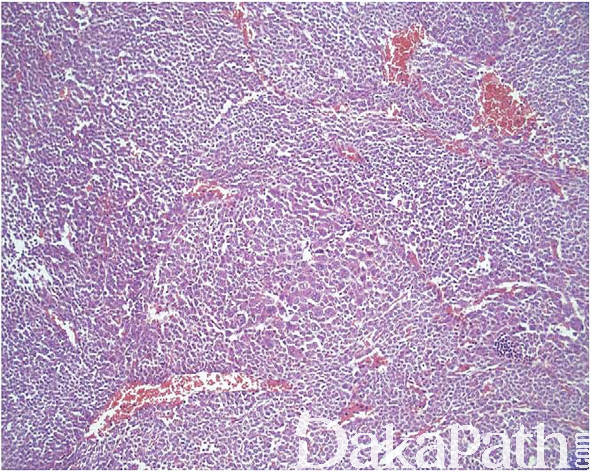
罕见;
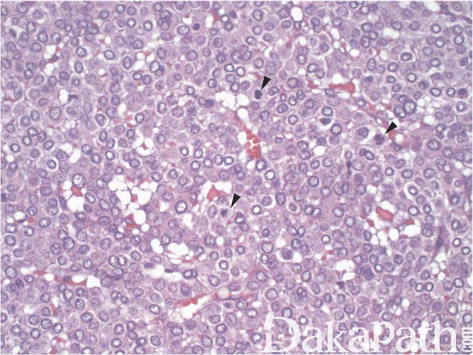
组织学上有两种类型:一种在形态上类似于平滑肌肉瘤或纤维肉瘤;另一种在总体结构上类似良性血管球瘤,瘤细胞由高度恶性的圆形细胞组成,肿瘤内常可见到良性血管球瘤区域;
恶性血管球瘤的诊断有以下 2 个标准:
弥漫的核多形性核+任何水平的核分裂象
或 2)可见非典型性核分裂像;
- 血管球瘤不存在上述诊断恶性的 2 个标准但存在除了核多形性之外的非典型特征者,诊断为恶性潜能未定的血管球瘤,包括:肿瘤大于 2cm, 部位较深,核分裂象活跃等。
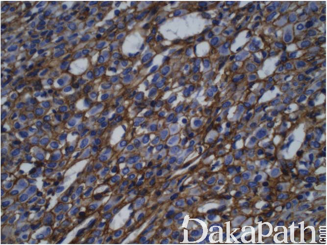
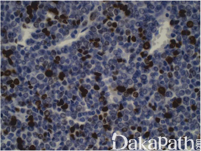
免疫组织化学染色:
在无良性血管球瘤背景的条件下,诊断恶性血管球瘤需要 α-SMA 和 Ⅳ 型胶原阳性,部分可表达 CD34 和 H-caldesmon,一般不表达 desmin
分子标记:
一部分显示恶性潜能未定或恶性特征的血管球瘤可见 BRAF V600E 突变
预后:
约 40%可出现转移和因疾病死亡
治疗:
完整切除
病例报道:
Lamba G, Rafiyath S M, Kaur H, et al. Malignant glomus tumor of kidney: the first reported case and review of literature [J]. Hum Pathol, 2011, 42(8):1200-3. 参考文献:
- Karamzadeh Dashti N, Bahrami A, Lee S J, et al. BRAF V600E mutations occur in a subset of glomus tumors, and are associated with malignant histologic characteristics [J]. Am J Surg Pathol, 2017, 41(11):1532-1541.
- Fletcher C D, Bridge J A, Hogondoorn P C, et al. WHO classification of tumours of soft tissue and bone [M]. Lyon: IRAC, 2013.
- Folpe A L, Fanburgsmith J C, Miettinen M, et al. Atypical and malignant glomus tumors: analysis of 52 cases, with a proposal for the reclassification of glomus tumors.[J]. American Journal of Surgical Pathology, 2001, 25(1):1-12.
← 恶性潜能未定的血管球瘤 软组织肌上皮瘤 →