睾丸恶性支持细胞瘤
Testicular Malignant Sertoli Cell Tumours
概述:
显示恶性组织学特点或生物学行为的睾丸支持细胞瘤
发病部位: 睾丸
诊断要点:
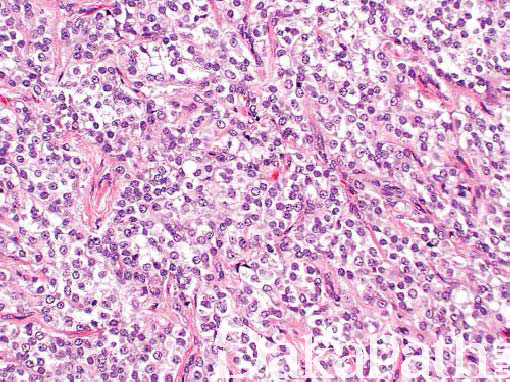
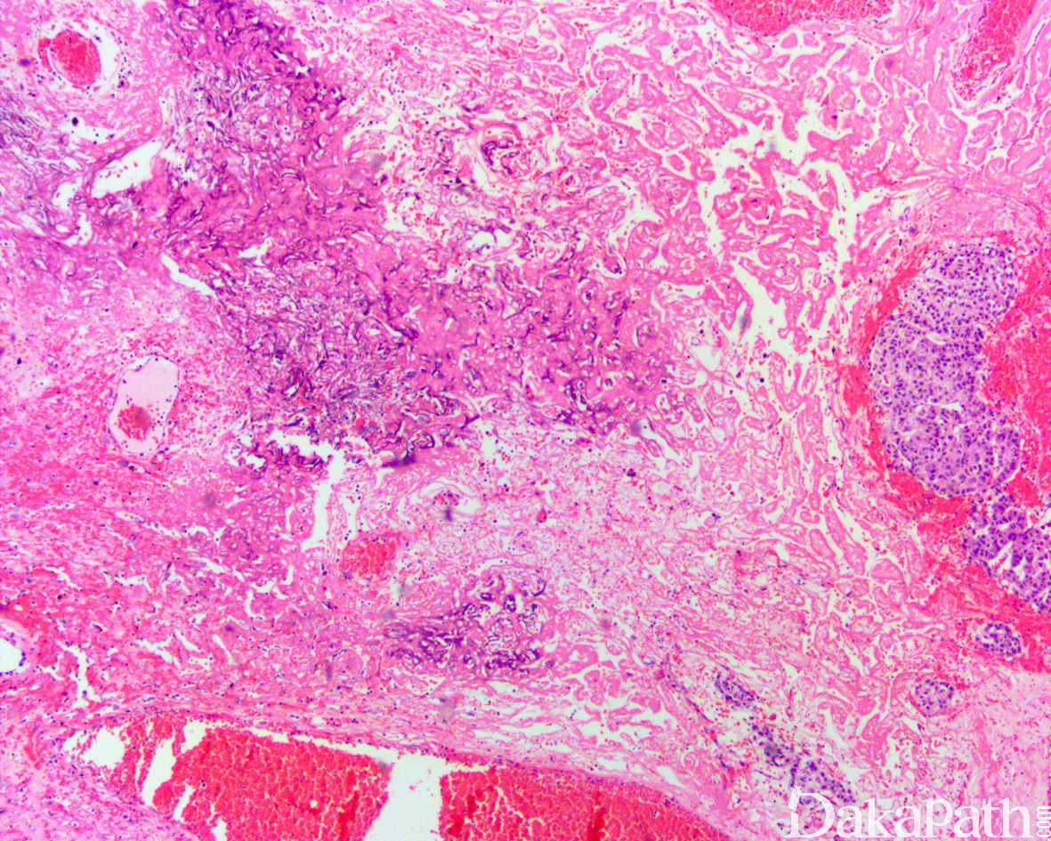
恶性 SCT 罕见,直径常>5cm(2 ~ 18cm),常见坏死和出血;
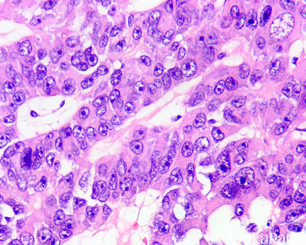
组织结构与细胞形态与良性者相似,以实性片状排列为主,细胞核多形,有一个或多个核仁,核分裂多见,常伴坏死;
纤维性间质、透明变性或黏液样间质不常见;
可见血管、淋巴管浸润;
可见数量不等淋巴浆细胞浸润,可有淋巴滤泡形成。
约 5%的 SCT,NOS 可表现为恶性的临床特征,ICD-O 编码为 3,组织学诊断恶性的标准主要有 5 项:瘤体直径> 5 cm,中-重度的细胞不典型性,核分裂象> 5/10 HPF,存在坏死,脉管侵犯等。Ulbright 和 Young 等指出,当存在上述 2 项或以上组织学特点时可诊断为恶性 SCT, 当仅有 1 项上述特点时宜归入“恶性潜能未定 SCT”范畴内,SCT,NOS 即使无任何上述特征,也不宜直接诊断为“良性 SCT”


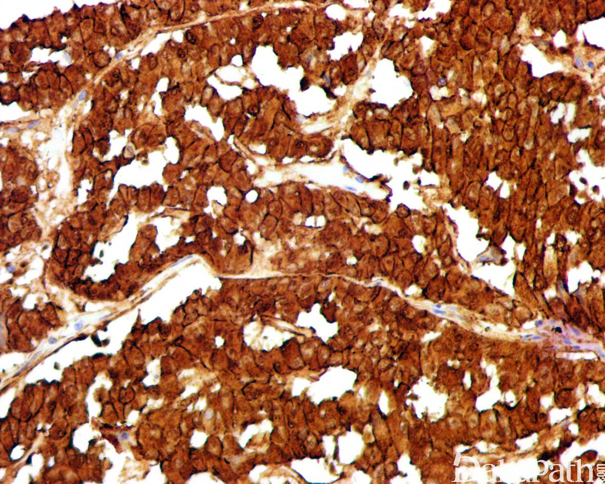
免疫组织化学染色:
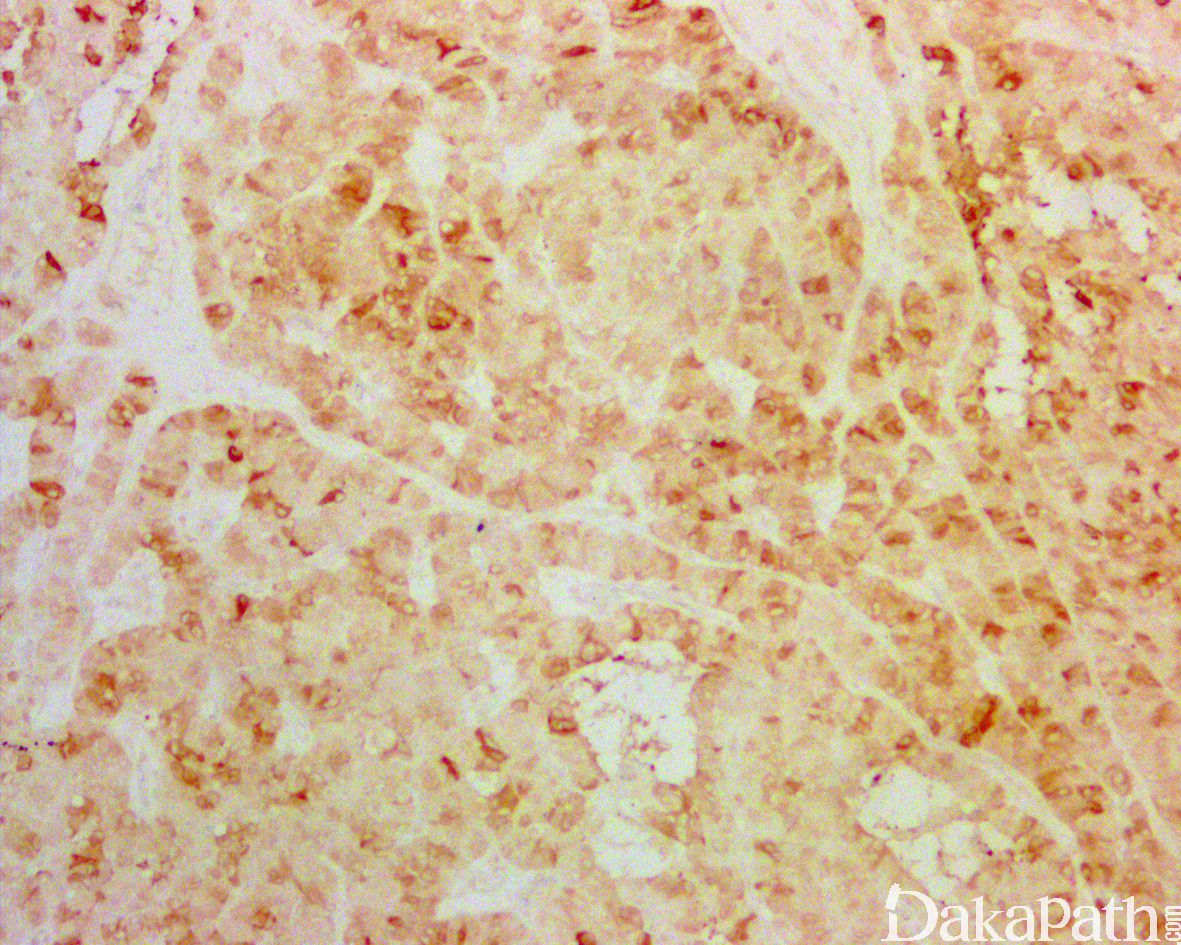
calretinin、SF1. 核 β-catenin、CD10. cylind1. CD99. melan A、WT-1. vimentin、S100 通常局灶或弥漫阳性,50%病例 inhibin 阳性,多数病例 CgA、Syn、AE1/AE3. 核 SOX9 阳性。
分子标记:
β-catenin 突变
鉴别诊断:
精原细胞瘤 :免疫组化染色表达 OCT3/4 和 SALL4。
卵黄囊瘤:免疫组化染色表达 GPC-3 和 SALL4。
精母细胞性肿瘤 :存在大中小三种类型的精母细胞。
治疗:
腹部放疗或化疗
病例报道:
Testicularsertoli cell tumoursand relative sub-types. Analysis of clinical and prognostic features.
参考文献:
Ulbright TM, Young RH. Tumors of the testis and adjacent structures, AFIP atlas of tumor pathology series 4[M]. Maryland: American Registry of Pathology, 2013. Young RH, Koelliker DD, Scully RE. Sertoli cell tumors of the testis, not otherwise specified: a clinicopathologic analysis of 60 cases [J]. Am J Surg Pathol, 1998, 22(6): 709-721. Henley JD, Young RH, Ulbright TM. Malignant Sertoli cell tumors of the testis: a study of 13 examples of a neoplasm frequently misinterpreted as seminoma [J]. Am J Surg Pathol, 2002, 26(5): 541-550.
← 睾丸间质细胞瘤 睾丸大细胞钙化性支持细胞瘤 →